Magnetic Resonance Imaging (MRI)
in Civil Engineering Fields
Kansai International Airport Land Development Co. Ltd
Intensities of magnetic resonance from nuclei of interest are recorded as a function of spatial positions tagged with pulsed field gradients. It is well known that physical characteristics of construction materials (e.g., clays, sands, and concretes) are significantly affected by water. It is why MRI has been anticipated to become a powerful tool of basic research in civil engineering fields. However, the use in the area has been limited so far and much remains to be explored.
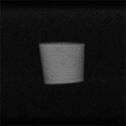
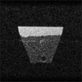
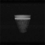
Fig. 1 2D Projection magnetic resonance image of water with white Portland cement (128*128).

Fig. 2 Digital photograph of the same sample.
Operator's Hand

Guu

Cyoki

Paa
TE=10ms TR = 500 ms NEX = 4 scans Total Imaging Time = 4 min 20 sec/ image
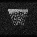
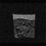
Glass Beads in Water
Digital Photo

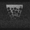
D ~ 12 mm
D ~ 6 mm
D ~ 2 mm
D ~ 1 mm

Digital Photo
Settled via gravity
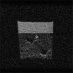
Consolidated
by hand-tapping
64th row of 128*128*128 data 3D spin-echo
TE =10 ms TR =100 ms NEX = 1 scan
Total Imaging Time = 12 sec/ image * 128 = 25 min
TE =10 ms TR =100 ms NEX = 1 scan
Total Imaging Time = 12 sec/ image * 128 = 25 min
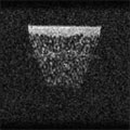
Bentonite and Water
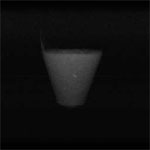
2D Projection

2D Slice
NEX = 32 scans TE = 10 ms TR = 100 ms
2D Spin-echo 128*128
2D Spin-echo 128*128
Masado Soil and Water
2D Projection